FIGURE 7.
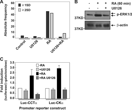
RA activates MAPK pathway in Neuro-2a cells. A, Neuro-2a cells were cultured in growing or differentiating media (containing RA 20 μm) in presence or absence of U0126 (10 μm) during 24 h and analyzed morphometrically. Graph depicts the absolute frequency of cells containing neurites equal to or longer than one soma diameter (≥1SD) or two soma diameters (≥2SD). B, Western blot analysis was used to investigate the phosphorylation state of ERK1/2 in Neuro-2a stimulated with RA (20 μm) during 1 h or without treatment. U0126 was used at 20 μm and preincubated during 15 min. C, Neuro-2a cells were transfected with Luc-CCTα and Luc-CKα promoter reporter construct. After 24 h, media were changed, and RA and U0126 (10 μm) were added to the indicated media and incubated 24 h. Luciferase and β-galactosidase activities were measured and data expressed as fold induction. The values are means ± S.E.

