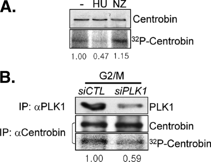
FIGURE 1.
PLK1 phosphorylation of centrobin at G2/M phase. A, HeLa cells were treated with 4 mm hydroxyurea (HU) or 0.33 μm nocodazole (NZ) for 24 h. During the last 3 h, the cells were cultured in the presence of [32P]orthophosphate and then subjected to immunoprecipitation followed by immunoblot analysis with the centrobin antibody. The amount of 32P-labeled centrobin was determined with autoradiography and measured densitometrically. B, the siPLK1-transfected HeLa cells were treated with 0.33 μm nocodazole for 16 h. During the last 3 h, the cells were cultured in the presence of [32P]orthophosphate. The endogenous centrobin and PLK1 proteins were immunoprecipitated (IP) with the specific antibodies and subjected to immunoblotting and autoradiography. The relative amount of [32P]centrobin was measured densitometrically.

