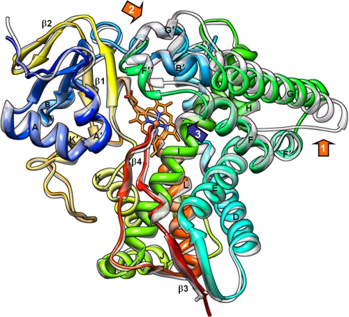
FIGURE 2.
Overall 14DM structure. Posaconazole-bound TC14DM (rainbow color, from blue N terminus to red C terminus) is superimposed with the ligand-free Tbb14DM (semitransparent gray); root mean square deviation for all Cα atoms is 0.91 Å. Major P450 structural elements are marked. The heme is shown in stick representation; posaconazole was deleted for clarity. The most flexible areas (GH loop and FG loop regions) are marked with orange arrows, 1 and 2, respectively. Repositioning the rigid I helix that is observed in all inhibitor bound trypanosomal 14DMs is marked with a blue arrow 3.

