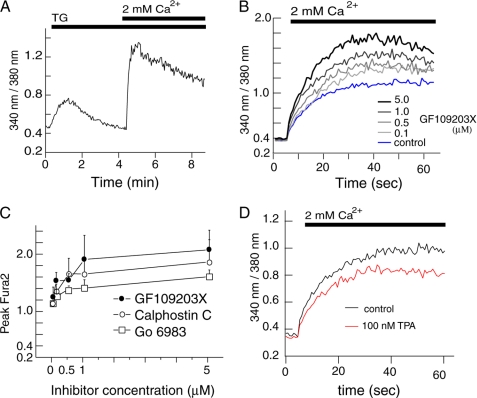
FIGURE 1.
PKC inhibition enhances SOCE in HEK293 cells. Intracellular Ca2+ concentrations were measured in HEK293 cells loaded with the Ca2+ indicator Fura2. A, to induce SOCE, ER Ca2+ stores were depleted with 1 μm thapsigargin (TG) in the absence of extracellular Ca2+. Subsequently, Ca2+ was added to a final concentration of 2 mm. Ca2+ levels are shown as the emission ratio of Fura2 following excitation at 340 and 380 nm. B, HEK293 cells were pretreated with different concentrations of GF109203X after depletion of Ca2+ stores with TG. Fura2 emission ratios were monitored for 60 s after addition of 2 mm Ca2+. C, peak values of Fura2 emission ratios obtained after preincubation of TG-treated cells with different concentrations of PKC inhibitors GFX (B), calphostin C (supplemental Fig. S1A) and Go 6983 (supplemental Fig. S1B) are shown as mean ± S.D. (n = 3). D, Fura2 emission ratios of TG-stimulated cells pretreated with 100 nm TPA followed by addition of 2 mm Ca2+.

