FIGURE 2.
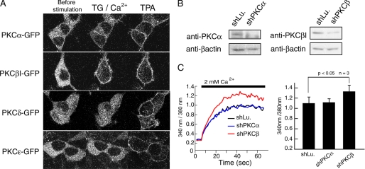
PKCβI but not PKCα negatively regulates SOCE. A, HEK293 cells were transfected with GFP-tagged PKC isoforms α, βI, δ, and ϵ. For confocal microscopy, cells were imaged before stimulation (left), after addition of TG in the presence of 2 mm Ca2+ (middle) and following stimulation with 500 μm TPA. B, for knockdown of PKCα and PKCβ, HEK293 cells were transfected with shPKCα and shPKCβ, respectively, and expression levels of PKCα and PKCβI were detected by Western blotting. Knockdown of luciferase (shLu) was used as negative control. β-Actin expression levels were used for normalization of protein expression. C, Ca2+ entry after pretreatment with TG was measured in HEK293 cells transfected with control and knockdown vectors as described in Fig. 1. Averages of peak Fura2 emission ratios after addition of 2 mm Ca2+ are shown (n = 3). Error bars, ±S.D.

