FIGURE 10.
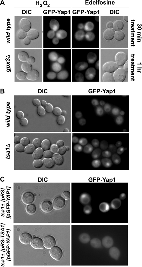
Constitutive nuclear localization of Yap1 in tsa1Δ cells. A, GFP fluorescence was monitored in gpx3Δ cells and their isogenic wild type (BY4741) expressing GFP-fused Yap1 as described above, after treatment with 0.5 mm H2O2 or 20 μg/ml of edelfosine (see “Experimental Procedures”). Nuclear translocation of Yap1 was evident within 30 min of treatment of wild type cells with either H2O2 or edelfosine, whereas 60 min were required to detect its nuclear localization in gpx3Δ cells treated with edelfosine. No Yap1 nuclear localization was detected in gpx3Δ cells treated with 0.5 mm H2O2 up to 1 h. B, GFP fluorescence was monitored in untreated tsa1Δ cells and their isogenic wild type (BY4741) expressing GFP-fused Yap1, or C, untreated tsa1Δ cells expressing GFP-fused Yap1 carrying an empty pRS316 vector or containing the TSA1 gene.

