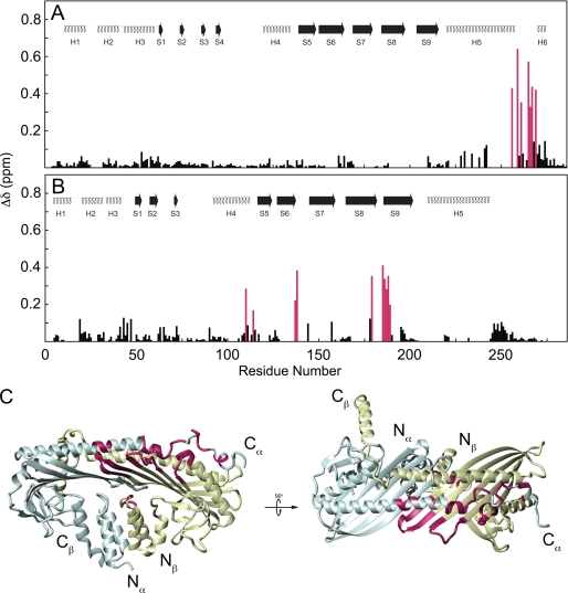
FIGURE 4.
Chemical shift map of V-1 binding on CP. Chemical shift change was plotted against residue number for CPα (A) and CPβ (B). Secondary structures are the same as in Fig. 1. Secondary shifts >0.15 ppm are highlighted purple. Residues experiencing large chemical shift changes were plotted onto the CP crystal structure (C). CPα is shaded blue; CPβ is shaded yellow. Residues whose chemical shift changed significantly upon V-1 binding are highlighted magenta. N and C termini of CPα and CPβ are indicated. Molecular structures were rendered using the program MolMol (64).

