FIGURE 2.
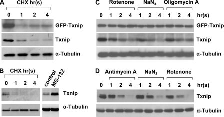
Decreased protein expression levels in cells treated by OXPHOS inhibitors. A, HeLa cells stably expressing a transgenic cytomegalovirus-driven GFP-Txnip fusion gene were treated with 40 μm cycloheximide (CHX) for the indicated times followed by immunoblot analysis of whole cell lysates. B, HEK293 cells were treated with 40 μm cycloheximide for the indicated times or with 20 μm proteasome inhibitor MG-132 for 12 h followed by analysis of endogenous Txnip expression. C and D, HeLa cells (C) and HEK293 cells (D) were treated with mitochondrial inhibitors for the indicated times followed by immunoblot analysis of whole cell lysates.

