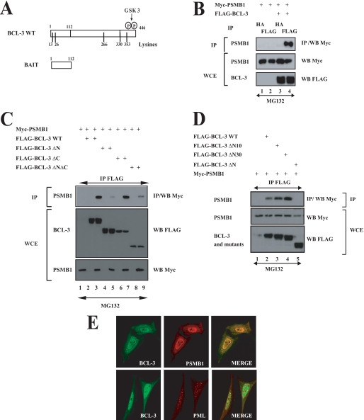
FIGURE 4.
PSMB1 is a BCL-3-interacting protein. A, a schematic representation of both BCL-3 and the bait used for yeast two-hybrid analyses. The GSK3 phosphorylation sites (Ser394 and Ser398) as well as the lysine residues (13, 26, 266, 330, and 353) on BCL-3 are illustrated. B–D, ectopically expressed BCL-3 and PSMB1 interact in mammalian cells through the N-terminal domain of BCL-3. 293 cells were transfected with the indicated expression plasmids. Cells were treated with MG132 (20 μm) for 4 h, and anti-HA (B) (negative control) or anti-FLAG (B–D) immunoprecipitations (IP) followed by an anti-Myc Western blot (WB) performed on the immunoprecipitates were carried out (top panels). Crude cell extracts were subjected to anti-Myc and anti-FLAG Western blots as well. E, PMSB1 and BCL-3 mainly co-localize in the nucleus. HeLa cells were transfected with FLAG-BCL-3 and with Myc-PMSB1, and their localizations were revealed through anti-FLAG and anti-Myc immunofluorescence, respectively. PML bodies also were visualized using the corresponding anti-PML antibody. WCE, whole cell extract.

