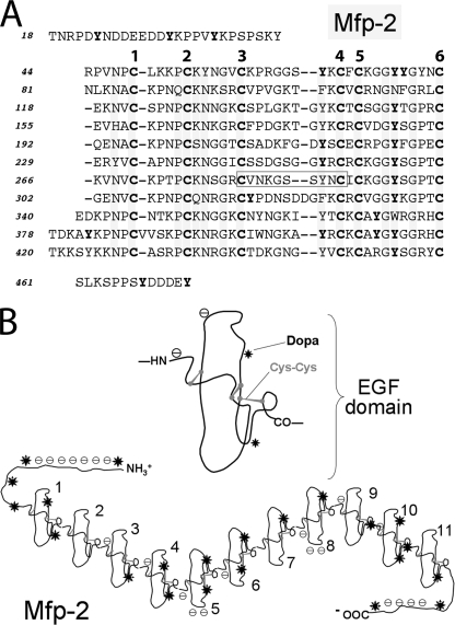
FIGURE 2.
Mfp-2. A, sequence is based on Mfp-2 from M. galloprovincialis (UniProtKB/Swiss-Prot entry Q25464) (13, 14). The 11 EGF repeats are aligned according to the invariant six cysteine residues/EGF. Tyrosine residues known to be occasionally or always modified to DOPA are denoted in bold. The boxed sequence in EGF repeat 7 is a calcium binding motif. Mfp-2 from M. edulis is 90% identical and is listed as (UniProtKB/Swiss-Prot entries Q1XBT6–Q1XBT8) but was never submitted as a published report. B, structure of one EGF domain based on Ref. 15, showing the cysteine residues paired for disulfides, charges, and location of the DOPA residues (*). Below the structure is a complete string of 11 EGF repeats showing the DOPA and acidic clusters.

