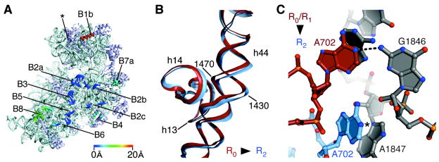
Fig. 3.
Contacts, or “bridges”, between the ribosomal subunits in the apo-70S ribosome in state R2. (A) The position of bridges in state R2 compared to those in state R0 (12, 13). Bridge numbering is the same as in (7). The direction of view and color coding are the same as in Fig. 2C. Bridge B1a (asterisk), includes the A-site Finger (H38 in 23S rRNA) which spans the subunit interface parallel to the A and P sites (29). This contact is not visible in the present structures due to disorder at the end of H38 in both states of the ribosome. (B) Bend in rRNA helix h44 in 16S rRNA that accommodates rotated state R2. Nucleotides and 16S rRNA helices are marked. The view is the same as in Fig. 2B. (C) Bridge B7a in state R2 compared to that in states R0 (12, 18–20) and R1 (14). Nucleotide A702 in 16S rRNA in the 30S subunit (light blue) and nucleotides in H68 of 23S rRNA in the 50S subunit (grey) are shown for state R2. Nucleotide A702 in state R0 or R1 is shown in red. The N1 position of A702 that would be methylated by dimethylsulfate is marked (5).

