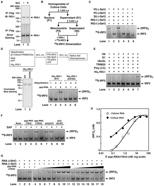
Figure 1. In Vitro Reconstitution of the RIG-I Pathway and Regulation of RIG-I by RNA- and Ubiquitination.
(A) Purification of RIG-I protein from Sendai virus-infected (+SeV) or untreated (−SeV) HEK293T cells stably expressing RIG-I containing a C-terminal Flag epitope. (B) Procedures for isolation of crude mitochondria (P5) and cytosol (S5) by differential centrifugation. (C) Virus-activated RIG-I induced IRF3 dimerization in vitro. The reconstitution reaction contained mitochondria (P5), RIG-I isolated from virus-infected or untreated cells, cytosolic extracts (S5) from uninfected cells, 35S-IRF3 and ATP. Dimerization of IRF3 was analyzed by native gel electrophoresis. (D) In vitro activation of RIG-I by 5′-pppRNA and ubiquitination. His6-tagged RIG-I (wild-type or WT) or its ATPase mutant (K270A) was purified from Sf9 cells (lower left panel), then incubated with 5′-pppRNA (79 nucleotides), ATP, and ubiquitination enzymes as outlined in the diagram. After incubation, aliquots of the reaction mixtures were further incubated with mitochondria (P5) and cytosol (S5) from uninfected cells together with 35S-IRF3 and ATP, and then IRF3 dimerization was analyzed by native gel electrophoresis. (E) In vitro activation of RIG-I by poly(I:C) and ubiquitination. Similar to (D), except that poly(I:C) was used and the dependency on ubiquitin and ubiquitination enzymes was tested. (F) The role of 5′-triphosphate for RNA to activate RIG-I in vitro. Similar to (D) except that the RNA was pretreated with or without shrimp alkaline phosphatase (SAP). (G) 5′-pppRNA and viral RNA are potent activators of the RIG-I pathway. Total RNA was extracted from HEK293T cells from viral-infected or untreated HEK293T cells, then incubated with RIG-I as in (D), followed by IRF3 dimerization assay (lanes 1–3). To measure the potency of 5′-pppRNA in RIG-I activation, increasing amounts of the RNA (135nt) (0.07 to 70 nM, at 3-fold increment) were incubated with RIG-I in the presence (lanes 12–18) or absence of cellular RNA from uninfected cells (lanes 5–11), then IRF3 dimerization assay was performed. (H) IRF3 dimer shown in (G) was quantified with ImageQuant, then plotted against the concentration of 5′-pppRNA. See also Figure S1.

