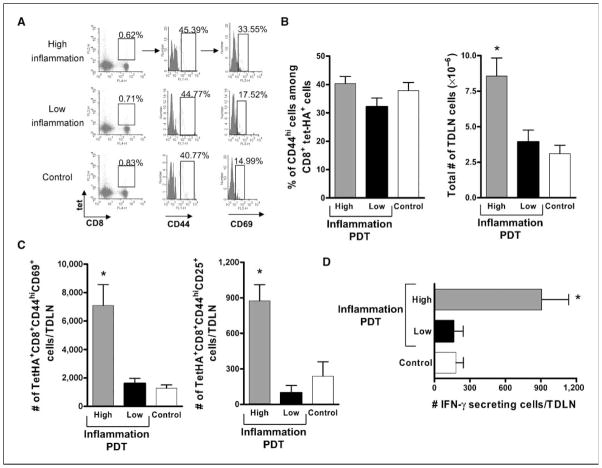
Figure 2.
Enhanced numbers of activated tumor-specific CD8+ T cells in TDLNs of mice following induction of acute local inflammation. BALB/c mice bearing Colo26-HA (A–C) or Colo26 (D) tumors were subjected to PDT treatments, causing different levels of acute inflammation, or injected with the photosensitizer and not exposed to laser treatment (Control). Twenty-four hours later, TDLN were harvested, single-cell suspensions generated, and the phenotype of CD8+ T cells specific for the HA antigen (tet+) was determined. A, flow cytometry depicting staining for tet-HA, CD8, CD44, and CD69. The boxed area in the first column identifies the tet-HA+CD8+ T cells. The second column depicts CD44 staining and is gated on the tet-HA+CD8+ T-cell population. The third column is gated on the tet-HA+CD8+CD44hi population and shows staining for CD69. The numbers indicate percentages of cells in the respective gates. Data from one representative experiment is shown. B, left, average percentage of CD44hi cells among the tet-HA+CD8+ population from three individual experiments. Right, the corresponding average number of live cells, as determined with trypan blue, recovered from the TDLNs. C, absolute number of tumor-specific CD8+ T-cell effector cells that expressed CD69 (left) and CD25 (right) following treatment. Experiments depicted in B and C were repeated twice with five mice per group; bars, SE. *, P < 0.03, compared with low-inflammation PDT. D, TDLNs were harvested at 24 h posttreatment and single-cell suspensions were generated. The number of cells able to specifically recognize tumor antigen AH1 and secrete IFN-γ as a consequence was determined with ELISPOT assays. Nonspecific staining with an irrelevant peptide (PA1) was subtracted from each group. Columns, average number of IFN-γ+ cells (each group contains at least six mice per group); bars, SE. *, P < 0.01, compared with low-inflammation PDT.

