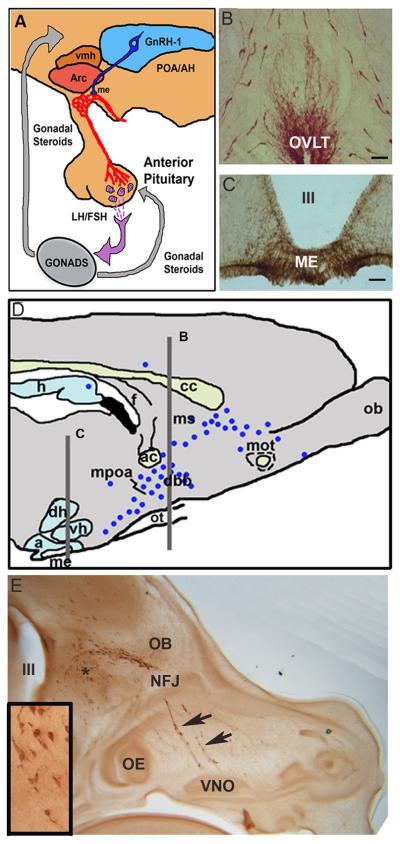
Figure 1. GnRH-1 neuroendocrine system.
A. Hypothalamic-Pituitary-Gonadal axis. GnRH-1 cells send axons to the median eminence (ME) where GnRH-1 is secreted into the portal capillary system. It is via this system that GnRH-1 cells affect gonadotropes of the anterior pituitary and subsequently gonadal function. POA/AH=preoptic area/anterior hypothalamus, vmh= ventromedial hypothalamus, arc=arcuate nucleus. C and D. Photomicrographs of sections immunocyto-chemically stained for GnRH-1 at the level of the OVLT (B) and ME (C). D. Parasagittal section of a rodent brain indicating the location of GnRH-1 cells (black dots, with levels in B and C shown by lines). GnRH-1 cells are distributed in a continuum from the olfactory bulbs to the caudal hypothalamus. h=hippocampus, f=fornix, ob=olfactory bulb, mot=medial olfactory tract, ms=medial septum, db=diagonal band of Broca, mpoa=medial preoptic area, d=dorsomedial hypothalamus, ot=optic tract, v=ventromedial hypothalamus, a=arcuate nucleus, me=median eminence. E. Parasagittal section of an E14.5 mouse embryo head immunocytochemically stained for GnRH-1. GnRH-1 cells can be found migrating from the VNO through the nasal forebrain junction (NFJ) into the forebrain. Asterisk shows location of inset, III= third ventricle.

