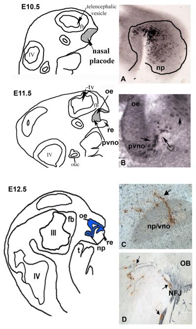
Figure 2. Development of the nasal placode/GnRH-1 neurons.
Camera lucida drawing from E10.5-E12.5 mice embryos showing invagination of the nasal placode and the development of the respiratory epithelium (re), olfactory epithelium (oe) and presumptive VNO (pvno)/nasal pit (np). tv=telencephalon, III=third ventricle, IV=fourth ventricle, t=tongue, fb=forebrain. A. Parasagittal section immunocytochemically stained for Hu (early neuronal marker) shows many positive cells within the nasal placode at E10.5. B. Parasagittal section from an E11.5 GnRH-GFP mouse immunocytochemically stained for GFP shows positive cells within the presumptive VNO (pvno) C. Parasagittal section from an E12.5 mouse, immunocytochemically stained for GnRH-1 (C) shows GnRH-1 cells leaving the VNO (arrows) D. Parasagittal section from an E12.5 mouse, immunocytochemically stained for GnRH-1 (brown) and peripherin (blue) shows GnRH-1 cells associated with peripherin fibers crossing the nasal forebrain junction (NFJ), peripherin axons entering the developing olfactory bulb (OB) and GnRH-1 cells still associated with a subset of peripherin axons turning caudally towards the hypothalamus.

