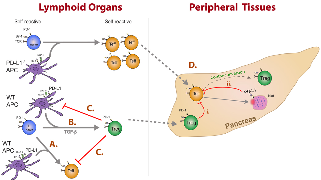
Fig. 6. The PD-1:PD-L pathway can regulate the dynamics between effector and regulatory T cells during multiple stages of autoimmune disease progression.
This figure illustrates consequences of these interactions in lymphoid organs and peripheral tissues. (A) PD-L1-expressing DCs stimulate PD-1 expression on naive T cells. PD-L1: PD-1 interactions limit T-effector cell expansion and survival. (B) In the presence of TGF-β, PD-L1-expressing DCs can promote the development of iTregs. (C) Tregs may restrain the magnitude of the immune response by inhibiting both DC and effector T-cell functions. (D) Activated T effector cells migrate to sites of inflammation where interaction with (i) PD-1-expressing Tregs or (ii) PD-L1 expressing target cells can (a) directly inhibit T-effector responses or may( b) facilitate the ‘contra-conversion’ of T effectors toward iTregs within the target organ. Additional cell types expressing PD-L1 such as the vascular endothelium and stromal cells may also influence T-effector cells but are not depicted in this figure for simplicity.

