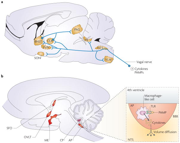
Figure 1. Pathways that transduce immune signals from the periphery to the brain.
The brain and the immune system communicate through different pathways. a | In the neural pathway, peripherally produced pathogen-associated molecular patterns (PAMPs) and cytokines activate primary afferent nerves, such as the vagal nerves during abdominal and visceral infections8,9 and the trigeminal nerves during oro-lingual infections10. Vagal afferents project to the nucleus tractus solitarius (NTS), and from there to the parabrachial nucleus (PB), the ventrolateral medulla (VLM), the hypothalamic paraventricular and supraoptic nuclei (PVN, SON), the central amygdala (CEA) and the bed nucleus of the stria terminalis (BNST). These last two structures form part of the extended amygdala, which projects to the periaqueductal grey (PAG). b | The humoral pathway involves circulating PAMPs that reach the brain at the level of the choroid plexus (CP) and the circumventricular organs11, including the median eminence (ME), organum vasculosum of the laminae terminalis (OVLT), area postrema (AP) and suprafornical organ (SFO). In the circumventricular organs, PAMPs induce the production and release of pro-inflammatory cytokines by macrophage-like cells expressing Toll-like receptors (TLRs). As the circumventricular organs lie outside the blood–brain barrier (BBB), these cytokines still need to reach the brain. They do so by mechanisms that are still unknown, but involve volume diffusion12.

