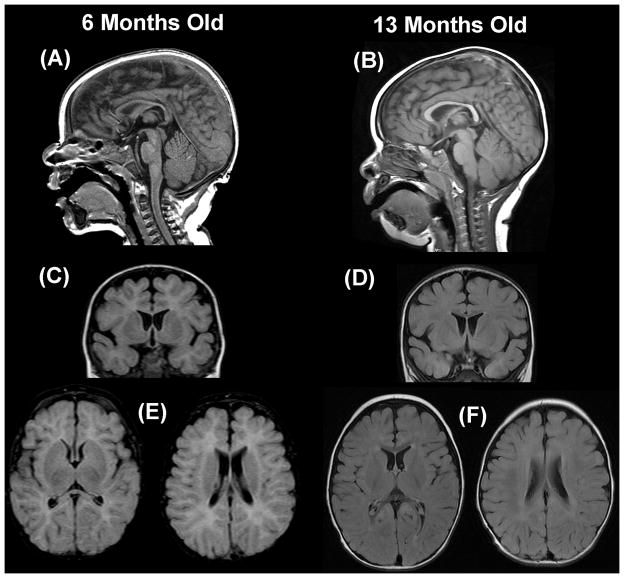
Figure 1.
T2 fluid attenuated inverse recovery magnetic resonance imaging of the patient’s brain at 6 and 15 months. Mid-sagittal images demonstrate that the corpus callosum is thin at 6 months (A) but a normal at 13 months (B). Excessive extra-axial space and underdevelopment of the operculum is seen on coronal and axial images at 6 months (C, E) but not 13 months (D, F).

