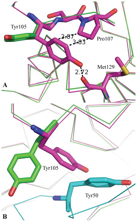
Figure 3.
A comparison of the Apo and bound structures of TEM. 3a: The green structure is that of TEM as bound to the BLIP from 1jtg70. The purple structure is that of apo TEM from the structure 1zg454. 3b: The green and purple structures are those of 3a. The cyan structure is that of BLIP from the complex with TEM. From 3b it is evident that Tyr105 of TEM-1 cannot adopt the same conformation in the bound (green) as observed in the apo (purple) due largely to the presence of Tyr50 of BLIP.

