Figure 3.
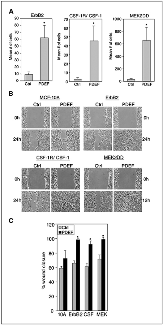
ErbB2, CSF-1R/CSF-1, and MEK2DD enhance PDEF induced migration. A, transwell migration assays of MCF-10A cells coexpressing ErbB2, CSF-1R/CSF-1, or MEK2DD with vector control or PDEF as described in Fig. 1B. Columns, mean number of motile cells per 20× field from six independent experiments; bars, SD. *, P < 0.005. B, wound healing assays of the same cell lines expressing vector control (Ctrl) or PDEF. Representative images captured with a 10× objective at the time of wounding (t = 0) and 12 hours (MEK2DD) or 24 hours after wounding. All experiments were repeated at least thrice. C, quantification of wound healing. Columns, percentage of wound closure at t = 24 hours [MCF-10A (10A), ErbB2, and CSF-1R/CSF-1 (CSF)] or t = 12 hours MEK2DD (MEK) averaged over four separate experiments (described in Material and Methods); bars, SD. *, P < 0.01.

