
Figure 3.
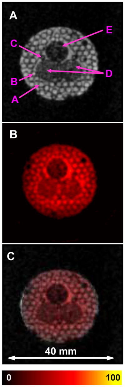
A. Middle slice of 3D proton NMR image; B. Corresponding slice of 3D EPR image. C. Superposition of A and B. Phantom was constructed from tubes containing 1 mM TAM. The phantom consisted of 20 mm long clusters of capillary tubes of 1.3 mm ID (A), 0.9 mm ID (B), 0.5 mm ID (C), 2 tubes of 6 mm ID with TAM surrounding voids from packed 0.4 mm OD rods (D), 1 tube of 6 mm ID with TAM surrounding voids from packed 0.3 mm OD rods (E). The central 3 large tubes were arranged into a triangular shaped pattern, and all were packed into an 18 mm i.d. plastic cylinder. Images of this phantom demonstrate good sensitivity and accurate co-registration for EPR and NMR images. The parameters used for the EPRI acquisition: frequency 1.1 GHz, microwave power ~20 mW, modulation amplitude 0.03 mT at 100 kHz, field gradient 0.1 T/m, projection scan time 1.3 sec, 46×46 projections FOV 32×32 mm. 3D MRI images were collected using a conventional gradient echo pulse sequence with the imaging parameters: 1H resonance frequency = 16.1805 MHz, spectral width = 10 kHz, TE/TR = 13/1000 ms, FOV = 32 mm, FOV in slice selection direction = 32 mm, Matrix size = 128 × 128 × 32, image orientation - axial, number of average = 1.