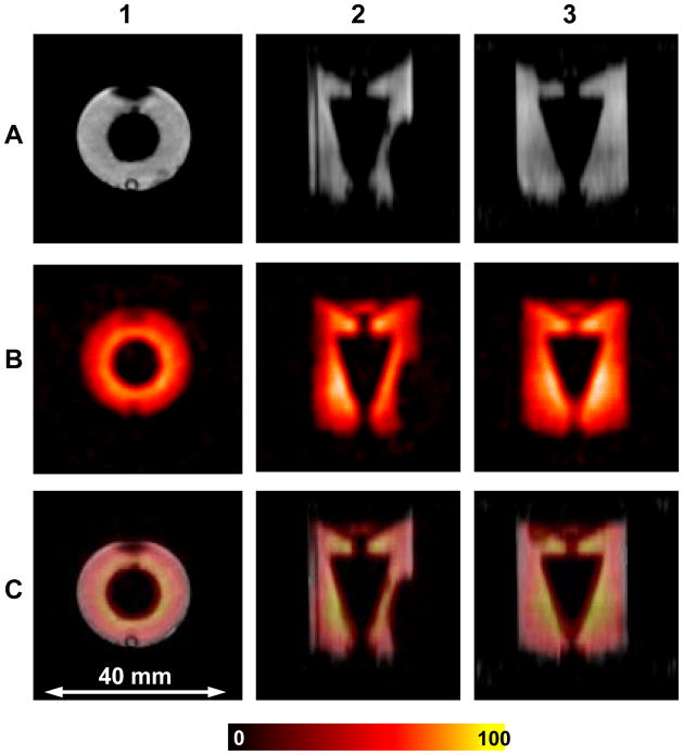
Figure 4.
Cylindrical phantom OD=19 mm with conical insert filled with 1 mM TAM. Row A shows MRI slices with axial (1), sagittal (2) and coronal (3) orientations. Row B shows the corresponding EPRI slices. Row C shows the superimposed images from A and B for each view. The parameters used for the EPRI acquisition: frequency 1.1 GHz, microwave power ~ 20 mW, modulation amplitude 0.03 mT at 100 kHz, field gradient 0.1 T/m, scan time 1.3 sec, FOV 32×32×32 mm 48×48 projections. The parameters for MRI: 1H resonance frequency = 16.1805 MHz, spectral width = 10 kHz, excitation sinc pulse (pulse length = 2 ms, bandwidth = 3 kHz) flip angle = 90°, TE/TR = 13/1000 ms, FOV = 32 mm, FOV in slice selection direction = 32 mm, image orientation - axial, matrix size = 128 × 128 × 32, number of average = 1.

