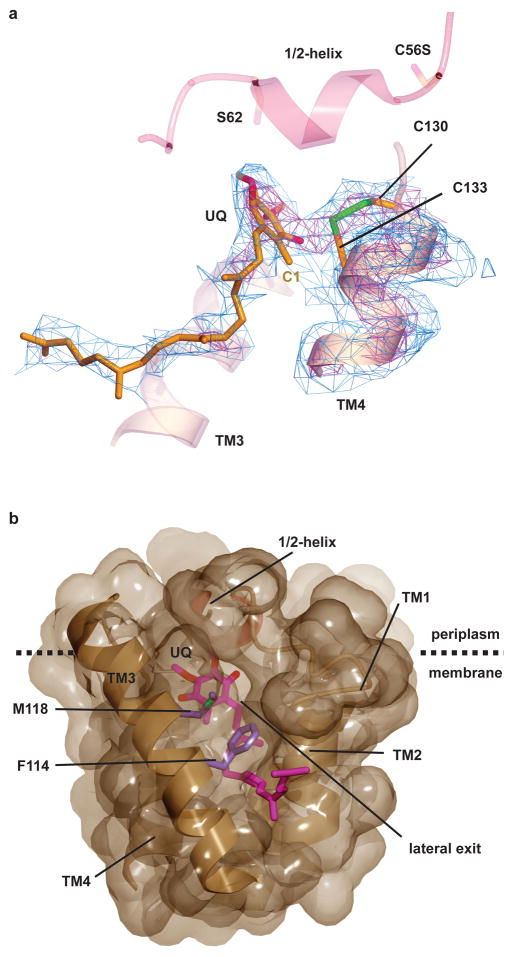
Figure 2. The active site of VKOR.
a, The active site of VKOR, including the cysteines of the CXXC motif in TM4 and the ubiquinone molecule (UQ). The experimental electron density map is contoured at 1σ (blue) and 3.5σ (red). Note the strong electron density between Cys130 and Cys133, indicative of a disulfide bridge (in green), as well as between Cys133 and the C1 position of the quinone. TM3 and the 1/2-helix are shown without electron density for orientation. b, Surface representation of the binding pocket for the quinone (in red), viewed from the side. TM5 was removed for clarity. Note the cleft between TMs 2 and 3, blocked only by the side chains shown in purple, which could provide a lateral exit for the quinone into lipid. The lower part of the membrane is not shown.

