
Figure 4.
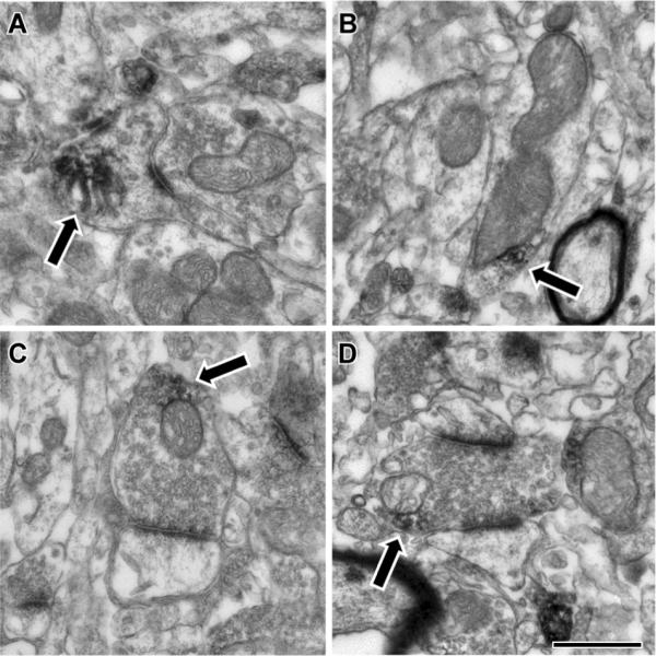
Electron microscopic localization of Cbln1 immunoreactivity in the rat striatum. Cbln1-ir (arrows) was found in many different subcellular compartments in the striatum, including axon terminals and dendrites. A) Within spines, Cbln1-ir was occasionally associated with the spine apparatus. B) In dendrites, Cbln1-ir was often observed adjacent to mitochondria, though other small vesicular structures were also seen nearby. C, D) Within axon terminals, Cbln1-ir was also often observed near mitochondria or vesicles in close proximity to the mitochondria. Synaptic contacts made by labeled terminals were always asymmetric and targeted both spines (panel C) and dendrites (D). Scale bar represents 500 nm in A, C, and D; and 600 nm in B.