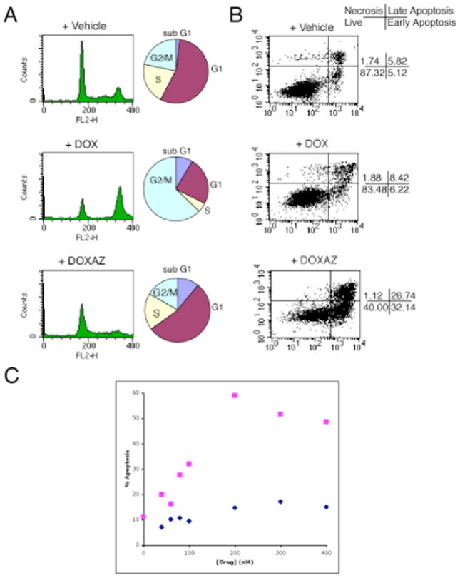
Figure 1.

Cell cycle distribution and apoptotic index analyses of HCT-116 cells exposed to doxorubicin or doxazolidine. HCT-116 cells were treated with 400 nM doxorubicin (DOX), doxazolidine (DOXAZ) or vehicle for 3 h and analyzed for (A) cell cycle distribution at 24 post-treatment and (B) apoptosis at 48 h post-treatment. (C) HCT-116 cells were treated with various concentrations of doxorubicin ( ) and doxazolidine (
 ) for 3 h and analyzed for apoptosis at 48 h post-treatment by annexin V staining.
