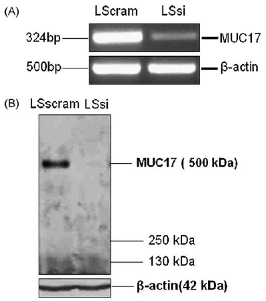
Fig. 2.

Expression analysis of MUC17. (A) RT-PCR for MUC17 mRNA expression in LSscram and LSsi cells. RT-PCR performed with B-actin primers were used to indicate mRNA in all lanes. (B) Western blot of cell lysates from LSscram and LSsi using anti-MUC17 antibody. Protein lysates from were resolved on 2% agarose gel containing SDS and transferred passively onto PVDF membrane. A unique band was revealed with a molecular weight consistent with the 500 kDa expected for MUC17. Loading control indicated by immunoreactivity using anti-actin antibody. Samples were tested in duplicate on two different occasions with similar results.
