Fig. 7.

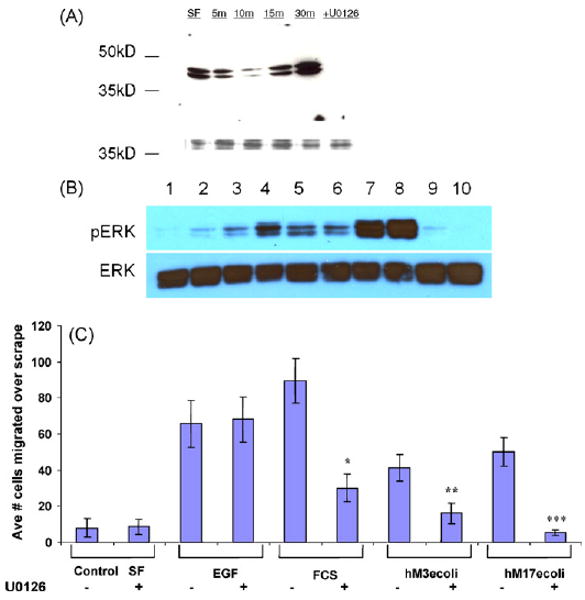
Activation of ERK cell signaling pathways. (A) IEC6 cells were treated with control serum-free medium (SF) or E. coli MUC17-CRD1-L-CRD2His8 (10 μg/ml) for 5–30 min and lysates were blotted with anti-phosphoERK1. Cells pretreated with ERK inhibitor U0126 before 15 min exposure to the MUC17 protein resulted in no phosphoERK1. The lower molecular weight bands indicate coumassie-stained protein demonstrating equal loading of gel. (B) YAMC cells were treated with control serum-free medium (SF. Lane 1); E. coli MUC17-CRD1-L-CRD2His8 (10 μg/ml) for 5, 10, 20, 60 or 120 min (lanes 2–6); EGF (100 ng/ml) for 5 and 10 min (lanes 7 and 8); and BSA (10 μg/ml) for 5 and 10 min; and lysates were blotted with anti-phosphoERK1. (C) Cell migration of Lovo cells treated with EGF (20 ng/ml), 10% fetal calf serum (FCS), Muc3-CRD1-L-CRD2 (hM3ecoli; 10 μg/ml), or MUC17-CRD1-L-CRD2 (hM17ecoli; 10 μg/ml); with or without pretreatment with 10 μM ERK inhibitor U0126 (u). *p < 0.01 vs. SF; **p < 0.01 vs. SF and p < 0.04 vs. hM3ecoli+u; ***p < 0.005 vs. SF and p < 0.02 vs. hM17ecoli+u. ERK inhibition has no effect on EGF-induced cell migration, partially blocks migration induced by 10% fetal calf serum (FCS), and completely blocks migration induced by both Muc3 and MUC17-CRD proteins.
