
Figure 2.
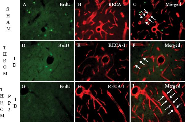

Intracerebroventricular (i.c.v.) injection of thrombin (20 U/animal, i.c.v.) causes reductions in brain microvascular endothelial cell (BMVEC) immunoreactivity after 1 day, and subsequent BMVEC proliferation around the rat brain vessels in lacunosum moleculare layer (LMol) of the hippocampus after 7 days and 14 days. Panels A-C show rats following sham operations labeled for BrdU, bromodeoxyuridine (A), RECA-1, rat endothelial antigen-1 (B) and the overlay or Merged image (C). RECA-1+ cells demonstrate the tube-shape of brain capillaries (arrows in panel C). Panels D to F show BrdU (D), RECA-1 (E) and the Merged image (F) at 1 day after thrombin injections. Compared with the sham group, RECA-1+ cells tend to lose their tube-shape at 1 day following thrombin injections (arrows in panel F) and there were no BrdU+ cells co-labeled with RECA-1 at one day (panel F). Panels G-I show the staining for BrdU (G), RECA-1 (H) and the Merged image (I) 1 day after thrombin plus PP2 injections. PP2 administration at day 0, immediately after thrombin injection, blocks the thrombin-induced loss of tube-shape of RECA-1+ cells. (Legend for first portion of Figure 2, Panels A-I).
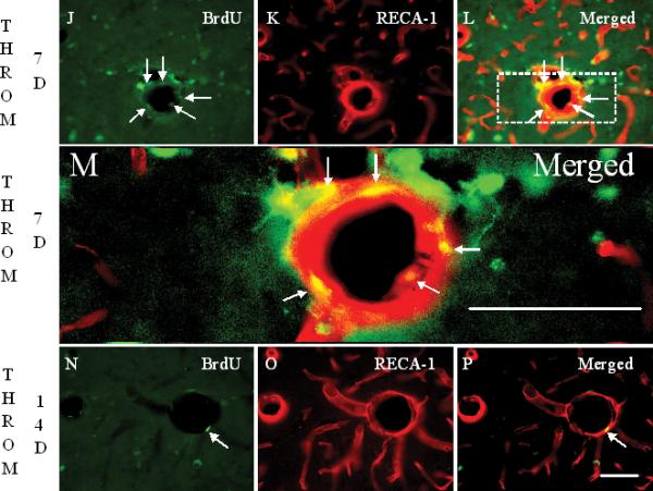
Panels J-L show the staining of BrdU (J), RECA-1 (K) and the Merged image (L) 7 days after thrombin injection. Compared to 1 day, BrdU+ cells are increased 7 days after thrombin injection. Some of these BrdU+ cells are co-labeled with RECA-1 (arrows in panel L and M). A few brain capillaries regained their tube-shape, though not completely. Panel M shows a higher power image of Panel L (area within dashed lines). RECA-1 stained BMVEC are red. The BrdU+/RECA-1+ double-labeled new born BMVEC nuclei are yellow. Panels N to P show the staining for BrdU (M), RECA-1 (N) and the Merged image (P) 14 days after thrombin injection. Compared to 7 days, BrdU+ cells are decreased, but some BrdU+ cells (N) remain co-labeled with RECA-1 (arrow in panel P), and more and more brain capillaries regained the tube-shape 14 days after the thrombin injection. Scale bars: A-P, 50 μm. (Legend for second portion of Figure 2, Panels J-P).
Panel Q shows the density of BrdU+ RECA-1+ double-labeled cells counted in LMol layer of hippocampus in each experimental group. Each column and vertical bar represents the mean ± standard error of the mean. ** p<0.01 vs. Cont (one-way ANOVA followed by Tukey's post hoc test). Cont = control. PP2 = non-specific src family kinase inhibitor. Throm = thrombin. RECA-1 = rat endothelial cell antigen-1. BrdU = bromodeoxyuridine, a marker of cell proliferation. (Legend for third portion of Figure 2, Panel Q).