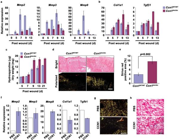
Figure 8. Enhanced fibrogenic response during wound healing in Ccn1dm/dm mice.
Gene expression in granulation tissues from WT and Ccn1dm/dm mice 5–13 days post-wounding was analyzed by qRT-PCR. Relative expression of (a) Mmp2, Mmp3, Mmp9, and (b) Col1a1 and Tgfβ1 is shown. (c) Total amount of hydroxyproline in healing wounds isolated 5–21 days post-wounding from WT and Ccn1dm/dm mice was determined and normalized over the total dry weight of the tissue. (d) Frozen sections from tissues 9 days post-wounding were stained with Sirius red for collagen. Images were acquired from bright field (top) and under polarized light (bottom). Scale bar = 100 μm. (e) Sirius red-positive area was calculated from at least 3 adjacent sections using ImageJ software. All experiments were done in triplicates and data presented as means ± S.D. (f) Purified recombinant CCN1 protein (0.1 mg/ml; 50 μl per dose) in PBS or PBS alone was topically applied to excisional wounds of Ccn1dm/dm mice daily, and wound granulation tissue harvested 9 days post wounding and analyzed by qRT-PCR (n=5). (g) Wounds so treated were sectioned and stained with Sirius red and visualized under polarized light (scale bar = 100 μm), and (h) stained for SA-β-gal (scale bar = 50 μm).

