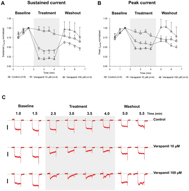
Figure 5. The phenylalkylamine voltage-operated Ca2+ channel blocker verapamil reversibly inhibits the inward current.
Both the (A) sustained and (B) peak components of the inward current are significantly inhibited by 10 µM and 100 µM verapamil. For each fiber tested, in control and test groups, peak and sustained currents were normalized to corresponding current values obtained at 1.5 minute after the start of the trial. Data were plotted as mean ± SEM. Seals and break-in were performed with cells constantly perifused with control solution. Cells remained under the perifusion for the whole length of the trial. The shaded box indicates time points where cells were exposed to the drug-containing bathing solution (for the test groups) or control solution itself (for the control group); (C) Representative traces of ICa/Ba activated by depolarizing S. mansoni muscle fibers from Vh of -70 mV to +20 mV with a 200 ms test pulse in the absence or presence of verapamil (10 µM or 100 µM). The dotted line denotes zero current level and the scale bar is 20 pA (traces from different fibers are scaled equally). The discontinuous time scale of the experiment is shown above traces, and each of the depolarizing pulses is 200 ms.

