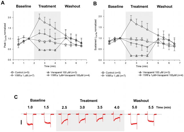
Figure 7. YIRFamide-mediated increase of ICa/Ba in S. mansoni isolated muscle fibers is reduced during treatment with the Ca2+ channel blocker verapamil.
Verapamil reduced the enhancement of ICa/Ba produced by YIRFamide, for both the (A) sustained and (B) peak currents. In each graph, data are compared showing currents under control conditions, the inhibition produced by 100 µM verapamil, the enhancement produced by 1 µM YIRFamide, and the co-application of both verapamil and YIRFamide, which in both cases most closely resembles the control currents. (C) Representative traces of ICa/Ba before, during and after treatment with 100 µM verapamil and 1 µM YIRFamide show currents that are effected by the co-application, but are intermediate to either agent applied alone. Data are plotted as mean ± SEM. Both verapamil and YIRFamide were dissolved in extracellular recording solution and delivered simultaneously during the time span indicated by a shaded box. The dotted line denotes zero current level and the scale bar is 20 pA. The discontinuous time scale of the experiment is shown above traces, and each of the depolarizing pulses is 200 ms.

