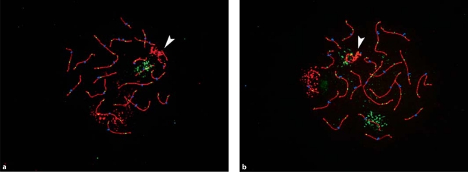
Fig. 1.
Representative images of rhesus male pachytene preparations. Antibodies detect the synaptonemal complex protein SYCP3 (in red) and the DNA mismatch repair protein MLH1 (in green) and CREST antiserum-positive signals (in blue) detect centromeric regions. (a) Rhesus chromosomes 1 (in red; homologous to HSA1) and 20 (in green; homologous to HSA16) were identified using FISH paint probes. (b) Rhesus chromosomes 4 (in red; homologous to HSA6) and 15 (homologous to HSA9) were similarly identified. In each image, an arrowhead denotes the XY bivalent.

