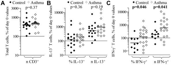
Figure 1.
CD3-stimulated accumulation of T cell subsets from control and asthmatic subjects. PBL from control (filled symbols) and atopic asthmatic (open symbols) subjects were cultured 5-d with CD3+CD28 monoclonal antibody + IL-2 + anti-IL-12, and then stimulated and analyzed for cytokine production by immunofluorescence-flow cytometry. Each symbol indicates the proportion (%) and number (n) of: (A) total; (B) IL-13+; and (C) IFN-γ+ T cells, expressed a percent of the respective day 0 values for each subject tested. Bars = geometric mean for each parameter. p-values from two-tailed student t-tests for differences in geometric mean between control and asthmatic subjects are shown.

