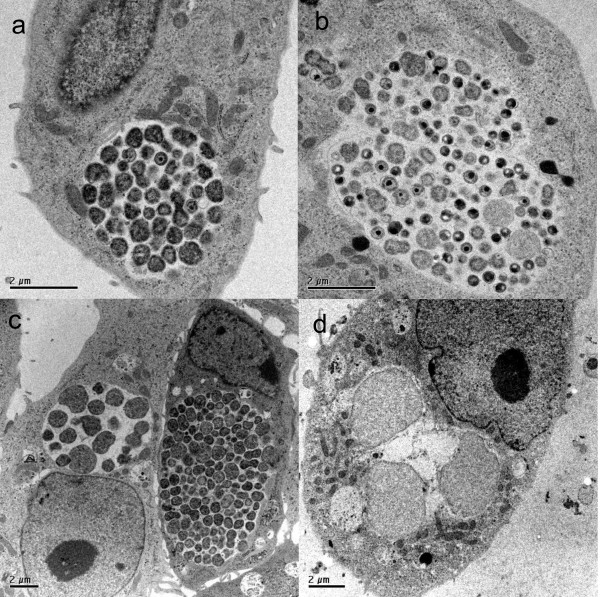
Figure 3.
Ultrastructure of chlamydial infection. Vero cells were infected with Chlamydia abortus (MOI 1) or Chlamydia pecorum (MOI 1), respectively for 39 h, fixed with glutaraldehyde, and further processed as described in material and methods. a) Chlamydia abortus mono infection containing many RBs and a few EBs. b) A more lobular Chlamydia pecorum mono infection inclusion containing many RBs, IBs and EBs. c) Chlamydia abortus double infection with ca-PEDV showing an inclusion of the growing phenotype on the right aspect of the picture and an inclusion consisting of RBs and large aberrant bodies in the adjacent cell on the left aspect of the picture. d) Chlamydia pecorum double infection with ca-PEDV depicting a small inclusion with aberrant bodies exclusively.

