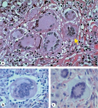
Fig. 2.
Histological images of multinucleated giant cells. a Langhans giant cells and one foreign-body giant cell (arrow) in a granuloma composed entirely of multinucleated giant cells. b Foreignbody giant cell. c Touton giant cell from a cutaneous juvenile xanthogranuloma. Images provided courtesy of Yale Rosen.

