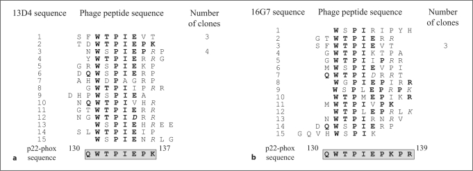
Fig. 3.
Phage display epitope mapping for mAb 13D4 (a) and mAb 16G7 (b). After selection of phages on an mAb 13D4 (a) or mAb 16G7 (b) affinity matrix, the nucleotide sequence was determined on isolated phage clones. The corresponding peptide sequences were aligned to identify a consensus sequence, which was compared to p22-phox sequence in order to identify the epitope region. Residues of phage peptides identical to residues of p22-phox are in bold. The epitope sequence is framed.

