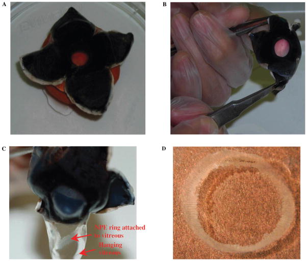
FIGURE 1.
Dissection technique of porcine eye to isolate intact NPE layer. (A) Cross section of the sclera at the posterior pole, which allows the eye to open by folding the resultant four flaps. (B) The technique of everting the cornea by pulling the cut flaps of the sclera backward and at the same time pushing the cornea inward with the index finger. (C) The method of separation of NPE layer along with vitreous. (D) The isolated intact ring of NPE layer. Please note that the NPE ring isolated from the porcine eye showed no detectable contamination by the PE or pigment granules.

