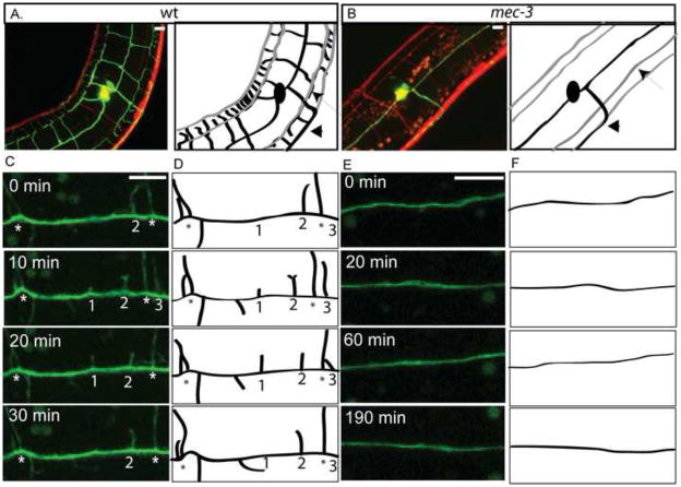
Figure 5. Dynamic initiation of PVD secondary branches is disrupted in mec-3 mutants.
Confocal images and schematic tracings of PVD::GFP (green/black) and panneural::dsRed (red/gray) (anterior left, ventral down) show that sub-lateral nerve cords (arrow) and PVD axon (arrowhead) are not altered in mec-3 mutants (B) in comparison to WT (A). Images (C) and schematics (D) from time-lapse confocal microscopy of wt L2 larval stage demonstrate dynamic PVD 2O branches (1–3) that initiate and retract in vicinity of established 2O branches (*) over 30 min period. Images (E) and schematics (F) of mec-3 mutants do not show PVD 2O branch initiation during 190 min of observation. Scale bar is 5 um. See supplemental movie 2 for wt and supplemental movie 7 for mec-3.

