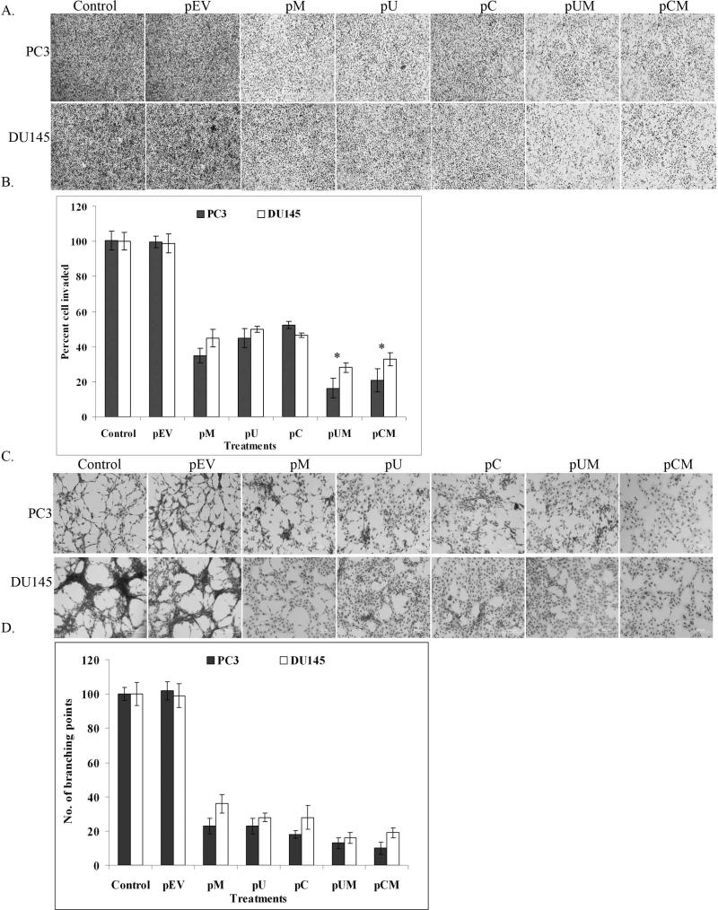
Figure 3. Down regulation of MMP-9, uPAR and cathepsin B reduced tumor invasion and angiogenesis.
(a) Matrigel invasion assay was performed after transfecting PC3 and DU145 cells with pM, pU, pC, pUM, pCM and pEV. After 48 h of transfection, the cells (1 × 10⁵) were allowed to invade through the transwell inserts coated with matrigel (1 mg/ml). After 18 h of incubation, the invaded cells were stained with Hema-3, photographed under light microscope and counted. (b) Invasion assay was quantified by counting the number of cells invaded through the matrigel in at least 5 representative fields. Bars represent the mean S.D from three different experiments, * (p < 0.01) represents difference between controls and pUM and pCM transfected cells. (c) In vitro angiogenesis assay was performed by incubating human microvascular endothelial cells in conditioned media collected from transfected and control cells. After 48 h incubation period the cells were stained with Hema-3 stain and photographed. (d) Angiogenesis was quantified by counting the number of branching points formed by HMEC grown in the cancer cell conditioned media with and without treatments. Values are mean S.D (* p<0.05) from three different experiments.

