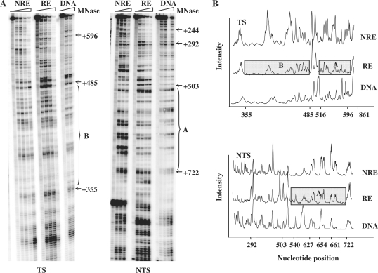
Figure 2.
MNase sensitivity of the chromatin containing the MseI restriction fragment of URA3 at the NRE and RE. (A) Typical footprinting autoradiographs showing MNase-sensitive sites in the TS and NTS of the MseI restriction fragment of URA3. DNA and chromatin samples from each strain (NRE and RE) were treated with increasing amount of MNase. Protected regions in chromatin at the repressive end are indicated by A and B. Nucleotide positions were indicated alongside the gels as numbers relative to the ATG start codon of URA3. (B) Relative MNase sensitivity after scanning the TS and NTS gels as shown in (A). The boxes indicate the regions that are less sensitive to MNase.

