
Figure 1.
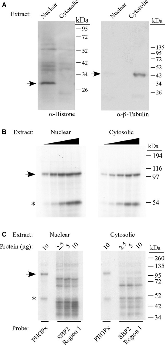
Identification of a 110 kDa SECIS-binding protein. (A) Nuclear and cytosolic extracts (30 µg protein) were analyzed by SDS–PAGE and western blotting using antibodies against histone H1 (nuclear marker) and β-tubulin (cytosolic marker) as indicated. The arrows indicate the positions of histone (left panel) and β-tubulin (right panel). (B) The 32P-labeled wild-type PHGPx SECIS probe was incubated with increasing amounts (2.5–20 µg) of nuclear or cytosolic extracts from McArdle 7777 cells in a UV crosslinking assay. The samples were analyzed by SDS–PAGE and autoradiography. The 110 kDa protein is indicated by the arrow. The band of ∼50 kDa, indicated by the asterisk, may represent a non-specific RNA-binding protein. (C) UV crosslinking experiments were performed with 2.5, 5 or 10 µg of nuclear or cytosolic extracts as described in (A) except that the 32P-labeled probe was either the PHGPx SECIS or an irrelevant RNA (region 1 from the SBP2 3′ UTR) that does not contain a SECIS but is predicted to form a stem-loop structure.