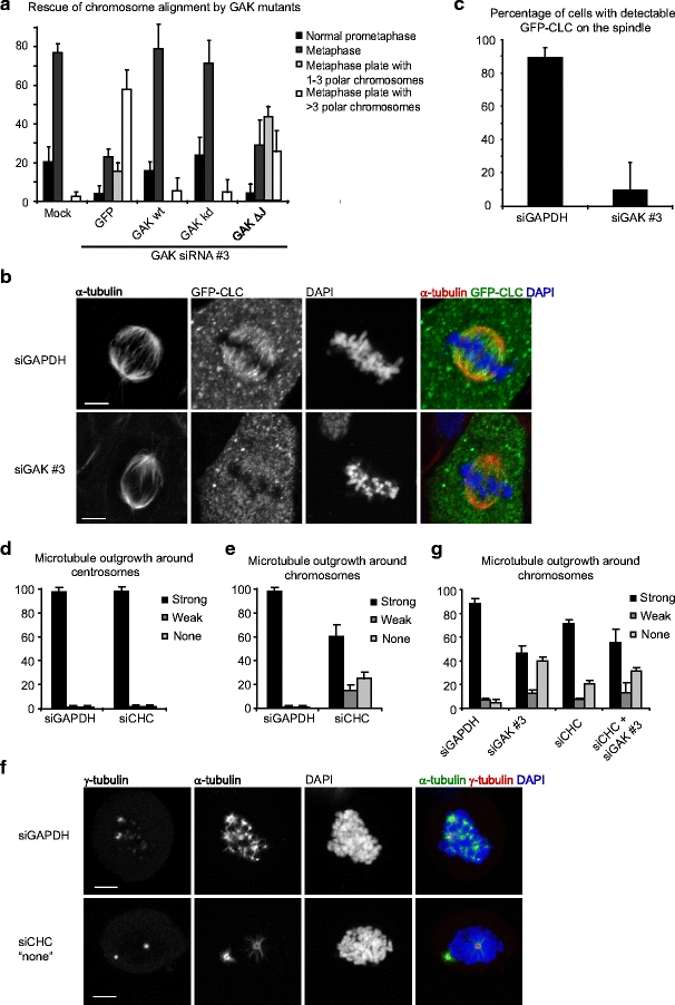
Fig. 4.

GAK mediates clathrin recruitment to the spindle. a Hela cells were transfected with indicated myc-tagged plasmids and 24 h later, cells were transfected with GAK siRNA where indicated. Forty-eight hours after siRNA transfection cells were fixed and stained for myc. DAPI was used to visualize the DNA. Chromosome alignment was then analyzed in mitotic cells expressing the indicated plasmids. b, c U2OS cells stably expressing green fluorescent protein (GFP)-clathrin light chain were transfected with indicated siRNAs. Twenty-four hours after transfection cells were re-transfected and 48 h after the first transfection cells were fixed and stained for α-tubulin and GFP. DAPI was used to visualize the DNA. a Representative cells. b Quantifications. d–g U2OS cells were transfected with indicated siRNAs. Twenty-four hours after transfection cells were re-transfected and 60 h after the first transfection cells were treated with nocodazole for 6 h, washed four times to remove nocodazole, fixed after 3 min, and stained for α-tubulin and γ-tubulin. DAPI was used to visualize the DNA. The amount of microtubules around centrosomes (visualized by γ-tubulin staining; d) or chromosomes (visualized by DAPI staining; e) was then analyzed. Images in (f) show a control cell (upper panel) and a clathrin heavy chain-depleted cell with a strong phenotype (lower panel). Graphs in (b–e, g) show averages of three independent experiments with 25 cells scored per condition per experiment in (c–e, g) and 50 cells per condition in (c). Error bars represent standard deviations. Scale bars in (a, f) indicate 5 µm
