
Fig. 4. Neurons release defective dG-VSV particles- ultrastructure.
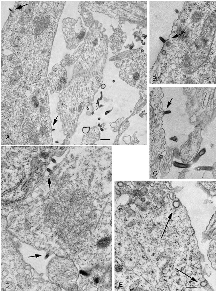
A. In this electron micrograph, several dG-VSV particles are seen budding off the dendritic or somatic membrane. To allow for a higher magnification image, cell body is cut off in the lower left part of the figure. B. Higher magnification of the particle budding from the proximal dendrite in the upper left part of A. C. Higher magnification of a virus budding from the membrane of the cell body, lower part of A. D. VSV buds off a glial cell. E. Although typical clathrin mediated endocytotic figures are seen, none contain newly generated virus particles. Bar, A, 350 nm; B-E, 225 nm.