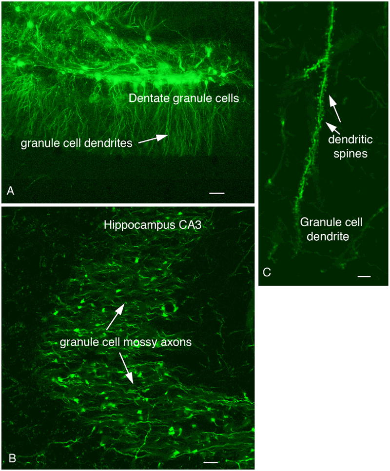
Fig. 5. Hippocampal mossy fibers and granule cell spines.

A. An injection of 100 nl of dG-VSV into the hippocampus dentate gyrus labels granule cells and dendrites. Scale bar, 35 um. B. Granule cell axons terminals, the mossy fibers, are labeled in CA3 after an injection of the dentate seen in A. Bar 6 um. C. Granule cell dendrites are covered with GFP-labeled spines. Bar, 5 um.
