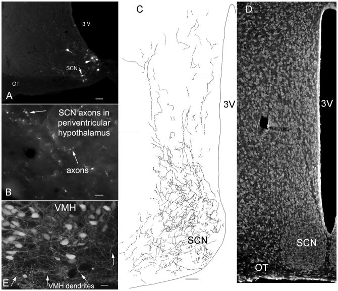
Fig. 7. Axonal projections of suprachiasmatic nucleus.
A. Injection of 50 nl of dG-VSV into the hypothalamic suprachiasmatic nucleus labels a few cells in the SCN. Bar, 50 um. B. In the area immediately dorsal to the SCN, axons and their boutons are labeled with GFP. Bar, 7 um. C. Camera lucida trace of GFP expressing axons within the SCN and in the area immediately dorsal to SCN. Bar, 40 um. D. Representative nissl-labeled section showing the SCN in parallel to C. E. Injections of dG-VSV into the ventromedial nucleus of the hypothalamus (VMH) showed that long dendrites (arrows) reached out of the core of the nucleus into the cell- poor shell. Bar, 12 um.

