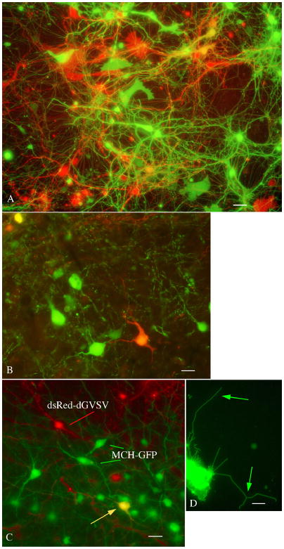
Fig. 8. Multicolor neuroanatomy with green and red dG-VSV.
A. In a culture from embryonic mouse brain, coinfection with green and red expressing dG-VSVs result in strong expression of green, red, orange, and yellow neurons and glia. This micrograph was recorded by sequential use of GFP and dsRed filter sets on the same field. Bar, 12 um. B. Similarly, when dG-VSV expressing red or green reporter genes are injected into the mouse cortex, within 20 hrs, green, orange, and yellow cells can be seen. Bar, 9 um. C. The dG-VSVdsRed can be coupled with expression of GFP in transgenic mice. Here, mice that express GFP in the hypothalamic neurons that contain melanin concentrating hormone were injected with the dG-VSVdsRed, resulting in one colabeled yellow cell, in addition to green MCH cells and red dG-VSV cells. Bar, 15 um. D. This micrograph shows a VSV-G protein directly labeled with GFP, allowing clear visualization of small and thin membrane bound cellular filopodia. Bar, 2.5 um.

