
Figure 2. Perivascular infiltration of cells into tumor tissues after treatment with 10 mg/kg/dose of anti-CTLA-4.
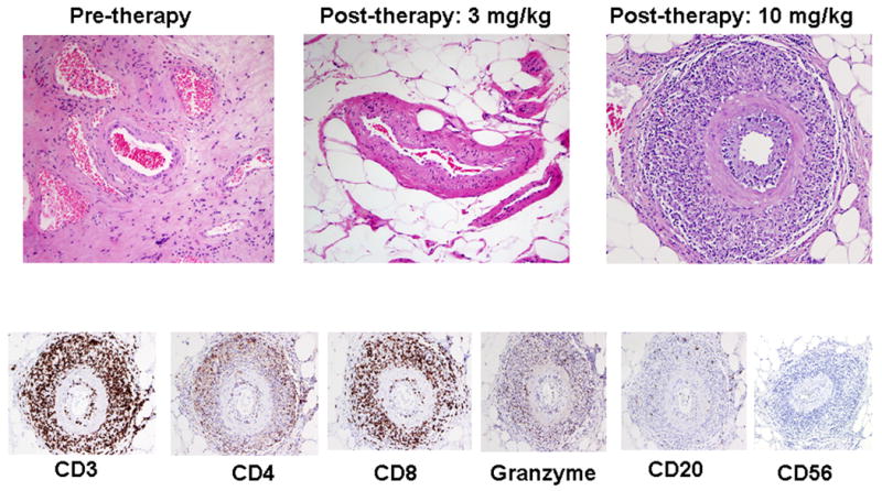
Representative pictures demonstrating an absence of perivascular infiltration of cells in untreated tumor tissues (0/11) and tumor tissues obtained from patients treated with 3 mg/kg/dose of anti-CTLA-4 (0/6) as compared to the presence of perivascular infiltration noted in tumor tissues obtained from patients treated with anti-CTLA-4 at 10 mg/kg/dose (2/5) (Upper Panel). Immunohistochemistry revealed that the infiltrating cells were positive for CD3, CD8, CD4 and granzyme but predominantly negative for CD20 and CD56 (Lower Panel).