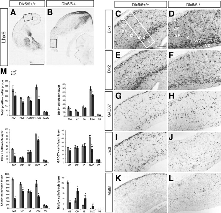
Figure 4.

A–M, Reduced number of Dlx1+, Dlx2+, GAD67+, and Lhx6+ cells in the lateral cortex of Dlx5/6−/− mutants at E16.5. in situ hybridization with probes for Lhx6 (A, B, I, J), Dlx1 (C, D), Dlx2 (E, F), GAD67 (G, H), and MafB (K, L) was performed on coronal sections of Dlx5/6+/ and Dlx5/6−/− mutants. The boxes in A and B show the region that is shown at higher magnification in C-L. The box in C shows the size of the region used for cell counting; the total number of cells expressing these genes within 125,000 μm2 of the lateral neocortex and the number of positive cells in each of the cortical layers are presented (M). The reduction is most severe in MZ and SVZ, particularly for Lhx6 and MafB within the MZ. Scale bars: (in A) A, B, 1 mm; (in C) C–L, 200 μm.
