Fig. 2.
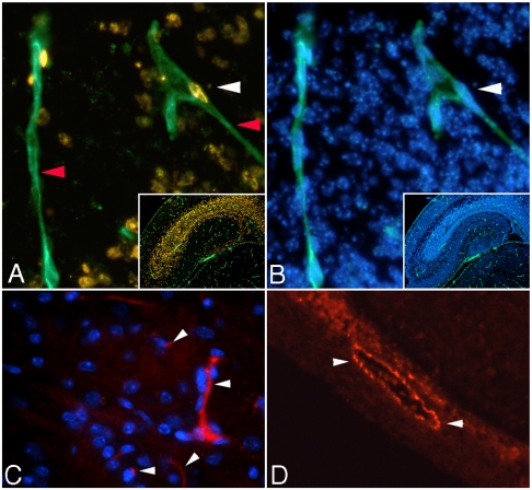
Immunohistochemistry studies in fetal brain. A and B: Representative 200X fluorescent images of the CORNUS AMMONI region 1 (CA1) of E17 hippocampus. A multiplex immunolabeling was performed for BrdU (golden staining—ALEXA 555, white arrowhead A), isolectin IB4 (green fluorescence—ALEXA 488, red arrowhead A) and DAPI (blue fluorescence when bound to genomic DNA, white arrowhead B). The proliferation index of EC was calculated as the ratio between the number of BrdU positive nuclei colocalized with isolectin (A) and DAPI positive nuclei colocalized with isolectin (B). Small insets are images of the entire hippocampus area captured with 50X magnification (A—isolectin-BrdU and B—DAPI-isolectin). C and D: Factor VIII RA staining of E17 fetal mouse hippocampus. Representative 200X fluorescent images of blood vessels expressing Factor VIII RA in a 5 μm hippocampal sections; image acquisition was performed with a rhodamine filter—blood cells fluoresce red (ALEXA 546 nm dye; white arrowheads) and nuclei fluoresce blue (DAPI staining). C shows small blood vessels with different trajectories and levels of Factor VIII as well as DAPI positive EC nuclei. D presents a more intense, Factor VIII positive, blood vessel with a bigger caliber situated in close proximity to the dentate gyrus.

