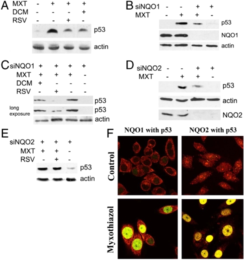
Fig. 5.
p53 induction in response to myxothiazol depends on NQO1 and NQO2. (A) Western analysis of p53 in RKO cells treated with 2 μM myxothiazol (MXT) for 10 h and 300 μM dicoumarol (DCM) for 4 h or 50 μM resveratrol (RSV) for 10 h. (B) Western analysis of p53 level in NQO1 knock-down (siNQO1) and control RKO cells after treatment with 2 μM myxothiazol for 10 h. (C) Western analysis of p53 in NQO1 knock-down RKO cells treated with 2 μM myxothiazol for 10 h, and 300 μM dicoumarol for 4 h or 50 μM resveratrol for 10 h. (D) Western analysis of p53 in NQO2 knock-down (siNQO2) and control RKO cells after treatment with 2 μM myxothiazol for 10 h. (E) Western analysis of p53 in NQO2 knock-down RKO cells treated with 2 μM myxothiazol and 50 μM resveratrol for 10 h. (F) Colocalization of p53 with NQO1 or with NQO2 after myxothiazol treatment. RKO cells were incubated with 2 μM myxothiazol or without (control) for 12 h and stained with antibodies to NQO2 or NQO1 (red) and p53 (green). Then pictures with red and green fluorescence were overlaid. Yellow color depicts colocalization of two proteins.

